IGF-1 LR3 1mg: The Growth Factor Research Peptide Explained

⚠️ Research Use Only. IGF-1 LR3 1mg is supplied strictly for in-vitro laboratory research. It is not intended for human consumption, therapeutic, diagnostic, or clinical use. All information below reflects peer-reviewed preclinical and scientific literature only. This content is not medical advice. Ascend Peptides UK accepts no liability for misuse of this product If you have spent any time exploring the world of peptide research, you will have come across the name IGF-1 LR3. It is one of the most studied and widely referenced growth factor peptides in the scientific community, used in laboratories across the UK and internationally to investigate everything from cell proliferation to metabolic signalling. This guide is designed as an educational resource for laboratory scientists, biotechnologists, and research professionals utilizing IGF-1 LR3 in formal, controlled in vitro studies. It outlines the biochemical structure, laboratory applications, and handling protocols for this widely referenced growth factor. Everything discussed here relates strictly to research and scientific investigation. Ascend Peptides UK supplies IGF-1 LR3 1mg and other high-purity research peptides exclusively for laboratory use, not for personal or clinical application. What Is IGF-1 and Why Is It Important in Research? To understand IGF-1 LR3, you first need to understand its parent molecule: insulin-like Growth Factor 1 (IGF-1). IGF-1 is a naturally occurring polypeptide hormone made up of 70 amino acids. While it is structurally similar to insulin and produced systemically by the liver (triggered by pituitary growth hormone signals), it is also synthesized locally in muscle, bone, and brain tissue, acting in both an autocrine and paracrine manner. In biological systems, IGF-1 plays a central role in regulating cell growth, differentiation, and survival. From a research perspective, IGF-1 sits at the crossroads of several major scientific fields: oncology, metabolic research, and intracellular signaling. Its receptor, the IGF-1 receptor (IGF-1R), is one of the most studied receptor tyrosine kinases in biomedical science. The downstream signaling cascades it activates, particularly the PI3K/Akt and MAPK/ERK pathways, govern everything from normal tissue growth to cancer cell survival. What Makes IGF-1 LR3 Different from Standard IGF-1? IGF-1 LR3 (Insulin-like Growth Factor 1 Long Arg3) is a synthetic, modified analog of IGF-1 specifically designed to perform more consistently in laboratory research conditions. It differs from native IGF-1 in two key structural ways: An arginine substitution at position 3 of the peptide chain (the source of “Arg3”). The addition of a 13-amino acid extension at the N-terminus, extending the total chain length from 70 to 83 amino acids. These modifications drastically alter how the compound behaves in experimental environments: Extended Half-Life & IGFBP Evasion: Native IGF-1 has a very short active half-life of approximately 12–15 minutes because it is rapidly sequestered by IGF-binding proteins (IGFBPs). The structural changes in LR3 dramatically reduce its affinity for IGFBPs, resulting in an extended in vitro half-life of roughly 20 to 30 hours. Clearance Mechanisms: Standard IGF-1 is also cleared via interaction with the IGF-2R/Mannose-6-phosphate receptor (M6PR). IGF-1 LR3 demonstrates reduced binding to this clearance receptor, further sustaining its bioavailability in experimental models. Receptor Nuance: While highly selective for IGF-1R, researchers must note that IGF-1 LR3 retains a residual affinity for the insulin receptor (IR) and hybrid IR/IGF-1R receptors, which is a critical variable when designing metabolic studies. Receptor Downregulation: Because of its extended half-life and continuous receptor stimulation, researchers running longer-duration assays must account for dose-dependent receptor downregulation as an experimental variable. Who Uses IGF-1 LR3 in Research and For What Purpose? IGF-1 LR3 is utilized across a broad range of scientific disciplines. Here are the main research contexts in which this compound plays a role: Cell Culture and Tissue Biology Research The most common application of IGF-1 LR3 in laboratory settings is as a defined growth factor supplement in cell culture media. Researchers working with serum-free or reduced-serum formulations use it to support consistent cell proliferation and viability. Its extended in vitro activity makes it exceptionally well-suited to longer-duration culture experiments where sustained receptor stimulation is required. Cancer and Oncology Research The IGF-1 signaling axis is extensively studied in cancer biology. IGF-1R overexpression or dysregulation has been observed in multiple cancer cell line models and is associated with tumor progression, resistance to targeted therapy, and dysregulated cell survival in multiple solid tumor models. Oncology researchers use IGF-1 LR3 to reliably activate IGF-1R signaling in these models, enabling the investigation of receptor inhibition strategies and drug resistance mechanisms. Metabolic and Endocrine Research Given the structural overlap between IGF-1 and insulin, IGF-1 LR3 is used in research exploring insulin signaling and metabolic regulation at the cellular level. Its distinct binding profile makes it a useful tool for dissecting the signaling crosstalk between these related pathways. Private and Independent Laboratories Beyond formal academic and institutional settings, IGF-1 LR3 is also utilized by private research entities and independent biochemistry laboratories. Understanding how this peptide influences cellular growth signaling remains a vital area of study in advancing in vitro methodologies. Ascend Peptides UK welcomes inquiries from all verified, legitimate research entities. How to Source IGF-1 LR3 1mg in the UK: Quality Indicators The quality of your research compound directly affects the reliability of your results. When sourcing an IGF-1 LR3 supplier, rigorous analytical standards are non-negotiable. Look for these key quality indicators: HPLC-Verified Purity: Purity should be confirmed via High-Performance Liquid Chromatography, ideally exceeding 98% for research-grade compounds. Mass Spectrometry (MS): Verifies that the peptide’s molecular weight and sequence precisely match the expected 83-amino acid structure. Endotoxin Testing (LAL Test): Crucial for cell culture applications. The compound must be tested via the Limulus Amebocyte Lysate (LAL) assay to ensure endotoxin levels are low enough that they will not trigger immune responses or compromise cell viability. Lyophilised Format: Freeze-dried peptides maintain stability far better than liquid formats during storage and transit. Batch Traceability: Essential for referencing specific production lots in published research and ensuring consistency across longitudinal experiments. At Ascend Peptides UK, our IGF-1 LR3 1mg meets all of these stringent standards. Whether you represent an academic institution, a biomanufacturing facility, or a private
CJC-1295 Without DAC: The Growth Hormone Research Peptide Explained

⚠️ Research Use Only. CJC-1295 Without DAC is supplied strictly for in-vitro laboratory research. It is not intended for human consumption, therapeutic, diagnostic, or clinical use. All information below reflects peer-reviewed preclinical and scientific literature only. This content is not medical advice. Ascend Peptides UK accepts no liability for misuse of this product If you work in peptide research or follow the science of growth hormone signalling, CJC-1295 without DAC is a name you will encounter frequently. It is one of the most studied growth hormone-releasing hormone (GHRH) analogues in the scientific community and for good reason. Its carefully engineered structure gives it distinct advantages over native GHRH in laboratory settings, making it a reliable and versatile tool for controlled research. This guide is provided by Ascend Peptides UK for laboratory scientists and research professionals. It offers a detailed biochemical overview of this compound, its pharmacological profile, and its applications in the study of the somatotropic axis. All information here relates strictly to scientific investigation. Ascend Peptides UK supplies this and other high-purity research peptides exclusively for laboratory use, not for human consumption, clinical application, or therapeutic purposes. Key Takeaways Scientific Identity: CJC-1295 Without DAC (frequently referred to as Modified GRF 1-29) is a synthetic, 29-amino-acid analogue of naturally occurring Growth Hormone-Releasing Hormone (GHRH). Structural Advantage: The peptide features four strategic amino acid substitutions that provide robust resistance to enzymatic breakdown by DPP-IV, significantly extending its stability in laboratory models compared to native GHRH. Release Profile: Unlike the DAC-bound variant, CJC-1295 Without DAC stimulates a time-limited, pulsatile release of growth hormone, making it the preferred reagent for studying natural, episodic secretion dynamics. Research Applications: It is heavily utilised in in vitro and animal model studies focusing on the hypothalamic-pituitary-somatotropic axis, IGF-1 pathways, and GHRH receptor binding kinetics. Handling and Storage: For optimal integrity, the lyophilised powder must be reconstituted with appropriate sterile solvents (such as bacteriostatic water) and stored in single-use aliquots at -20°C to avoid damaging freeze-thaw cycles. Strict Regulatory Status: This compound is an experimental research chemical. It is not FDA or MHRA approved and is strictly prohibited for human consumption, therapeutic application, or clinical use. Understanding Growth Hormone-Releasing Hormone — The Scientific Foundation To appreciate the significance of this compound, it helps to first understand the molecule it is designed to mimic: Growth Hormone Releasing Hormone, or GHRH. GHRH is a naturally occurring peptide hormone produced in the hypothalamus. Its primary role is to stimulate the anterior pituitary gland to synthesise and secrete growth hormone (GH). This process forms part of the hypothalamic-pituitary-somatotropic axis, one of the most studied regulatory systems in endocrine biology. Growth hormone itself has wide-ranging downstream effects across biological systems, influencing cell growth, metabolism, body composition, and the production of Insulin-like Growth Factor 1 (IGF-1) in the liver. Understanding how GHRH drives GH secretion and how this process can be reliably modelled in research settings is the scientific foundation on which this research peptide category is built. Native GHRH has a significant limitation as a laboratory tool: it is rapidly degraded by a serum enzyme called dipeptidyl aminopeptidase IV (DPP-IV), giving it a very short half-life in biological environments. This makes sustained research into its effects difficult. That is precisely the problem that synthetic GHRH analogues were developed to address. What Is CJC-1295 Without DAC? CJC-1295 Without DAC, also referred to as Modified GRF(1-29) or Mod GRF 1-29 is a synthetic analogue of the first 29 amino acids of GHRH, which is the biologically active portion of the molecule. The ‘Without DAC’ designation is important: it distinguishes this compound from its related counterpart, CJC-1295 With DAC, which uses a different modification to extend its half-life. The modifications incorporated into this peptide are four strategic amino acid substitutions at specific positions in the chain. These substitutions achieve two research-relevant goals: Increased resistance to DPP-IV degradation, the primary enzyme responsible for breaking down native GHRH in biological environments Improved receptor binding stability, enhancing the compound’s potency and consistency in controlled laboratory assay conditions The result is a growth hormone research peptide that retains the functional characteristics of native GHRH whilst offering significantly improved in vitro stability — making it a far more practical tool for systematic laboratory investigation of the GHRH-GH signalling axis. CJC-1295 Without DAC vs CJC-1295 With DAC: Understanding the Difference This is one of the most common questions in this research area, and understanding the distinction is essential when selecting the right compound for a given experimental design. The key difference lies in the presence or absence of a Drug Affinity Complex (DAC), a modification that enables the peptide to bind covalently to albumin in the bloodstream, dramatically extending its half-life. CJC-1295 without DAC produces a pulsatile pattern of GH stimulation in research models. Each administration generates a defined, time-limited pulse of GH receptor activation closely reflecting the natural episodic GH release seen in biological systems. This makes it particularly suited to research examining pulsatile GH secretion dynamics. CJC-1295 with DAC binds to albumin via the DAC technology, producing sustained, continuous GH pathway activation over an extended period. This is preferred for studies requiring prolonged GH stimulation rather than pulsatile release. For researchers studying natural GH physiology and pulsatile secretion, the Without DAC variant is generally the more appropriate research compound. Both are available from Ascend Peptides UK, and selecting between them depends entirely on the experimental design and research objectives. Research Applications in Laboratory Settings This peptide has been the subject of a substantial body of published research. The following summarises the primary laboratory contexts in which it is applied: 1. Growth Hormone Axis Research The most direct application is the investigation of the GHRH-GH signalling axis itself. Researchers studying hypothalamic-pituitary function and GH secretion dynamics use this compound to reliably stimulate GHRH receptor activity in controlled experimental models, enabling systematic investigation of the somatotropic axis. 2. IGF-1 Pathway Studies Growth hormone stimulates the liver to produce IGF-1, making GHRH analogue research indirectly relevant to IGF-1 pathway investigation as well.
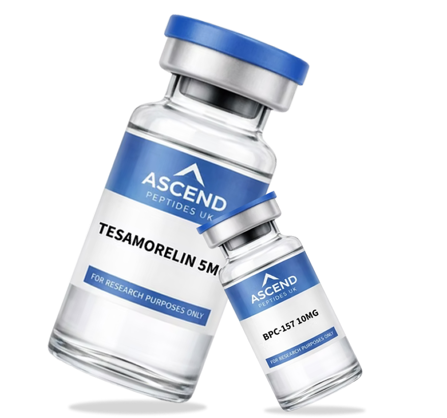
BPC-157 10mg Research Peptide: A Complete Laboratory Guide

⚠️ Research Use Only. BPC-157 10mg is supplied strictly for in-vitro laboratory research. It is not intended for human consumption, therapeutic, diagnostic, or clinical use. All information below reflects peer-reviewed preclinical and scientific literature only. This content is not medical advice. Ascend Peptides UK accepts no liability for misuse of this product Few research peptides have generated as much consistent scientific attention as BPC-157. Derived from a partial sequence of Body Protection Compound, a protein found naturally in gastric juice, this synthetic pentadecapeptide has been the subject of a substantial body of animal and in vitro laboratory research across a wide range of scientific disciplines. This guide is written for both laboratory scientists conducting formal research programmes and scientifically legitimate individuals who want to understand what this compound is, how it behaves in laboratory models, and why it has maintained such strong interest within the global peptide research community. Ascend Peptides UK supplies BPC-157 10mg as a high-purity research peptide for laboratory investigation only. All information in this guide is provided strictly for scientific and educational purposes. This compound is not intended for human consumption, clinical use, or therapeutic application. What Is BPC-157? Structure and Origin Explained BPC-157 is a synthetic pentadecapeptide, a chain of exactly 15 amino acids. The name stands for Body Protection Compound 157, referring to a specific experimentally designated sequence derived from the parent protein during research development. The numbering reflects its place in a series of sequences tested from the parent BPC protein, rather than a positional reference within the protein chain itself. That parent protein, Body Protection Compound, is found naturally in human gastric juice, where it plays a reasonably well-established role in gastric cytoprotection. The 15-amino acid sequence of this compound is: Gly-Glu-Pro-Pro-Pro-Gly-Lys-Pro-Ala-Asp-Asp-Ala-Gly-Leu-Val, with a molecular weight of approximately 1,419 Daltons. This particular arrangement gives the peptide its distinctive biochemical properties and distinguishes it from other synthetic research peptides in terms of its stability profile and interactions in laboratory models. One of the most scientifically interesting characteristics of this compound is its notable stability in biological environments. Unlike many peptides that degrade rapidly when exposed to gastric acid or enzymatic activity, this sequence demonstrates remarkable resistance to hydrolysis under acidic conditions, a property that has made it an interesting subject for laboratory research and a useful tool for studying peptide stability across a range of experimental conditions. Mechanism of Action: What Laboratory Research Has Found The precise mechanism by which BPC-157 exerts its effects in laboratory models is an active area of scientific investigation. It is worth noting upfront that no specific receptor for BPC-157 has been conclusively identified to date; this is an important caveat for researchers designing receptor-binding studies, as it distinguishes this compound from peptides with a well-defined single-receptor mechanism. Instead, published laboratory research has documented interactions across multiple molecular pathways. These vary in the strength and consistency of supporting evidence, so it is useful to distinguish between the better-established findings and those representing a more preliminary evidence base. More consistently replicated findings: Nitric oxide (NO) system modulation: The most robustly documented mechanism across published studies. Multiple independent research groups have identified interactions between this peptide and the nitric oxide pathway, which plays a central role in vascular biology, inflammation signalling, and cellular communication. Growth factor upregulation (VEGF pathway): Also well-represented in the published literature. Laboratory research has documented interactions with vascular endothelial growth factor (VEGF) and related growth factors involved in angiogenesis, the formation of new blood vessels, making this a significant area of interest in regenerative research. More preliminary findings, documented but less consistently replicated: FAK-paxillin pathway: Research has identified activation of the focal adhesion kinase (FAK) and paxillin signalling pathway, involved in cell migration and tissue organisation in laboratory models. The evidence base here is smaller and warrants cautious interpretation. Neurotransmitter system interactions: Published research has examined interactions with dopaminergic, serotonergic, and GABAergic systems in animal models, contributing to its use in neurobiological research contexts. These findings are documented but derive predominantly from animal studies and should be interpreted accordingly. Tendon and ligament fibroblast activity: In vitro studies have examined how this compound affects fibroblast behaviour, the cells responsible for producing collagen and extracellular matrix components, making it relevant to connective tissue research. Replication across independent research groups remains more limited in this area. It is important to note that the majority of this mechanistic research has been conducted in cell culture and animal models, with a significant proportion originating from a single research group. The compound is supplied strictly for laboratory research purposes, and none of this research constitutes clinical evidence of efficacy in humans. Laboratory Research Applications of BPC-157 10mg BPC-157 10mg has been applied across a broad range of laboratory research disciplines. The following summarises the primary areas in which this compound has been investigated in controlled scientific settings: 1. Connective Tissue and Musculoskeletal Research One of the most extensively published research areas involves this peptide in the context of connective tissue biology. In vitro and in vivo laboratory studies have examined its interactions with fibroblasts, tendon cells, and ligament tissue in controlled research models. Researchers investigating extracellular matrix dynamics, collagen synthesis, and fibroblast migration have used this compound to study cellular behaviour under controlled conditions. 2. Gastrointestinal Biology Research Given its origin from a gastric protein, gastrointestinal biology is one of the most natural research contexts for this compound. Laboratory studies have examined its interactions with gastric mucosal cells, intestinal epithelial biology, and inflammatory signalling in gut tissue models. Researchers studying mucosal integrity, gastric ulcer models, and intestinal permeability have used it to investigate cellular protection mechanisms in laboratory settings. 3. Angiogenesis and Vascular Research Research into the compound’s interactions with the VEGF pathway and nitric oxide system has made it a subject of interest in angiogenesis research, the scientific study of new blood vessel formation. Laboratories investigating vascular biology, wound healing models, and endothelial cell behaviour have incorporated this peptide into their research designs to examine how growth factor signalling influences
GHK-CU 50mg Research Peptide: A Complete Laboratory Guide

⚠️ Research Use Only. GHK-CU 50mg is supplied strictly for in-vitro laboratory research. It is not intended for human consumption, therapeutic, diagnostic, or clinical use. All information below reflects peer-reviewed preclinical and scientific literature only. This content is not medical advice. Ascend Peptides UK accepts no liability for misuse of this product GHK-CU is one of the most scientifically studied copper-binding peptides in the field of biochemical research. First isolated from human plasma in the early 1970s, this naturally occurring tripeptide-copper complex has since been the subject of an extensive body of published laboratory research spanning skin biology, wound-healing models, anti-inflammatory signalling, and gene-expression studies. This guide is written for two audiences. For laboratory scientists and research professionals, it provides a thorough scientific overview of this compound — its structure, mechanism of action, and applications in controlled research settings. For scientifically engaged individuals (qualified researchers, laboratory professionals, and academic or institutional research settings) who follow developments in peptide biochemistry and skin research science, it offers an accessible explanation of why this copper peptide complex has attracted such sustained scientific attention. Ascend Peptides UK supplies GHK-CU 50mg as a high-purity research peptide for laboratory investigation only. All content in this guide is provided strictly for scientific and educational purposes. This compound is not intended for human consumption, clinical use, or therapeutic application of any kind. What Is GHK-CU? Structure, Origin, and Unique Properties GHK-CU is a naturally occurring tripeptide-copper complex found in human plasma, saliva, and urine. The peptide component GHK consists of three amino acids: glycine (Gly), histidine (His), and lysine (Lys). The CU suffix denotes its association with a copper (II) ion, which the peptide binds with high affinity through the imidazole group of histidine and the amino-terminal amine. This copper-binding characteristic is central to the compound’s biochemical identity. Copper is an essential trace mineral involved in a wide range of enzymatic processes in biological systems, including collagen synthesis, superoxide dismutase activity, and angiogenesis. The ability of the GHK tripeptide to chelate and transport copper ions in laboratory models has made it a subject of considerable research interest in fields ranging from dermatology research to wound biology and gene regulation studies. What sets this compound apart from many other research peptides is its notably broad influence on cellular behaviour in laboratory models. Published research has documented interactions with over 4,000 human genes, a finding that has positioned this tripeptide as one of the most far-reaching small peptide sequences studied in modern biochemical research. It is this breadth of documented laboratory activity that makes it particularly relevant across multiple research disciplines. Mechanism of Action: How GHK-CU Behaves in Laboratory Models GHK-CU interacts with biological systems through several interconnected mechanisms, many of which have been documented across a substantial body of peer-reviewed research. Understanding these mechanisms is important for researchers designing experiments that incorporate this copper peptide complex as a research tool. Key mechanisms identified in published laboratory research include: Copper chaperone activity: This compound acts as a copper transport molecule in research models, delivering Cu(II) ions to copper-dependent enzymes, including lysyl oxidase (involved in collagen and elastin crosslinking) and superoxide dismutase (a key antioxidant enzyme). This copper delivery function is central to many of the biological activities observed in laboratory settings. Collagen and extracellular matrix regulation: Multiple in vitro studies have documented the influence of this peptide on fibroblast gene expression, collagen synthesis, and extracellular matrix remodelling. This has made it a widely used research tool in skin biology and connective tissue research. Antioxidant and anti-inflammatory signalling: Published research has shown interactions with antioxidant defence systems and inflammatory signalling pathways in laboratory models, including modulation of pro-inflammatory cytokine activity and upregulation of antioxidant enzyme expression. Angiogenesis research: Studies have documented the ability of this compound to stimulate angiogenic signalling in laboratory models, particularly through interactions with VEGF and FGF pathway components, making it relevant to wound biology and vascular research. Gene expression modulation: Genome-wide studies have identified interactions with thousands of human genes, including those involved in tissue remodelling, inflammation regulation, stem cell biology, and metabolic processes, highlighting the compound’s unusually broad influence in laboratory research contexts. These mechanisms have been studied predominantly in in vitro cell culture models and animal research models. This compound is supplied strictly for research purposes, and none of this research constitutes clinical evidence of efficacy in human subjects. Research Applications of GHK-CU 50mg in Laboratory Settings The breadth of published research on this compound reflects its versatility as a laboratory research tool. Here are the primary disciplines in which it has been applied in controlled scientific settings: 1. Skin Biology and Dermatological Research Skin biology is the most extensively published research area for this copper peptide complex. In vitro studies have examined its interactions with keratinocytes, fibroblasts, and melanocytes, the three primary cell types of the skin. Research in this field has focused on fibroblast proliferation and collagen gene expression, extracellular matrix dynamics, basement membrane integrity, and cellular responses to oxidative stress. For researchers investigating skin biology at the cellular and molecular level, this compound is one of the most well-characterised tools available. 2. Wound Healing Biology Research The compound’s documented interactions with fibroblast activity, angiogenic signalling, and extracellular matrix remodelling have made it a research tool of significant interest in wound healing biology. Laboratory studies have examined how this peptide influences fibroblast migration, collagen deposition patterns, and vascular endothelial cell behaviour in wound model systems. This research area has attracted sustained scientific attention and continues to generate active laboratory investigation. 3. Anti-Inflammatory and Antioxidant Research Published laboratory research has documented this compound’s interactions with inflammatory cytokine signalling, including modulation of TNF-alpha, IL-6, and other pro-inflammatory mediators in cell culture models. Its upregulation of antioxidant enzyme expression, including superoxide dismutase and catalase, has also been a subject of research interest. These properties have positioned it as a valuable tool in studies examining oxidative stress responses and inflammation-related signalling at the cellular level. 4. Hair Follicle Biology Research A notable area of published
Tesamorelin 10mg Research Peptide: A Complete Laboratory Guide

⚠️ Research Use Only. Tesamorelin 10mg is supplied strictly for in-vitro laboratory research. It is not intended for human consumption, therapeutic, diagnostic, or clinical use. All information below reflects peer-reviewed preclinical and scientific literature only. This content is not medical advice. Ascend Peptides UK accepts no liability for misuse of this product Tesamorelin is a synthetic analogue of Growth Hormone Releasing Hormone (GHRH) that has attracted significant scientific interest within the peptide research community. Unlike many GHRH analogues, which are based on shortened sequences of the endogenous hormone, this compound is a full-length GHRH analogue modified with a specific trans-3-hexenoic acid group at its N-terminus to improve stability and resistance to enzymatic degradation. This guide is designed for both laboratory scientists and research professionals seeking a thorough scientific overview of this compound, and for scientifically curious individuals (laboratory researchers, academic investigators, and scientific professionals) who want to understand what makes it unique among GHRH research peptides. Whether you are sourcing for an established research programme or exploring this compound for the first time, this guide covers everything you need to know. All information here is provided strictly for scientific and educational purposes. Ascend Peptides UK supplies this and other research compounds for laboratory investigation only, not for human consumption, clinical application, or therapeutic use of any kind. What Is Tesamorelin? Structure and Scientific Background Tesamorelin is a stabilised synthetic analogue of endogenous GHRH. Unlike shorter GHRH analogues such as Sermorelin (which replicates only the first 29 amino acids of GHRH) or Modified GRF 1-29, this compound replicates the full 44-amino acid sequence of native GHRH with a single but significant structural modification: the addition of a trans-3-hexenoic acid group covalently bonded to the N-terminal tyrosine residue. This modification is the defining structural feature of the compound and the source of its research significance. The trans-3-hexenoic acid group substantially increases resistance to DPP-IV (dipeptidyl aminopeptidase IV) enzymatic degradation — the primary enzyme responsible for the rapid breakdown of native GHRH in biological environments. As a result, this GHRH analogue maintains its receptor binding activity for considerably longer in research conditions than unmodified GHRH. The full 44-amino acid sequence also distinguishes this compound from shorter GHRH research peptides. Because it retains the complete GHRH sequence, it interacts with the GHRH receptor (GHRHR) in a manner that more closely reflects the endogenous ligand, making it a valuable tool for research requiring greater physiological relevance than truncated GHRH analogues can provide. Tesamorelin vs Other GHRH Research Peptides: Key Differences Understanding how tesamorelin compares to other GHRH analogues helps researchers select the most appropriate compound for their specific experimental design. Here is how this compound sits within the broader GHRH research peptide landscape: CJC-1295 Without DAC (Modified GRF 1-29) is based on the first 29 amino acids of GHRH with four stabilising amino acid substitutions. It produces pulsatile GH stimulation in research models and has a shorter sequence than this compound. For a detailed overview of that peptide, see our CJC-1295 Without DAC research guide. Tesamorelin, by contrast, uses the full 44-amino acid GHRH sequence with N-terminal modification, offering a different structural profile for researchers requiring full-length GHRH receptor engagement. Sermorelin is the unmodified GHRH(1-29) sequence, retaining structural closeness to native GHRH but with greater susceptibility to DPP-IV degradation. This compound’s N-terminal modification addresses this limitation whilst also preserving the complete GHRH receptor interaction surface. Downstream of the GHRH-GH axis, growth hormone stimulates hepatic IGF-1 production. Researchers studying the full length of this signalling cascade from GHRH receptor activation through to IGF-1 pathway effects may combine GHRH analogue research with IGF-1 receptor studies. Our IGF-1 LR3 1mg research guide provides a detailed overview of how IGF-1 LR3 functions as a research tool for investigating the downstream growth factor signalling pathway. Mechanism of Action in Laboratory Research Models Tesamorelin acts as a selective GHRH receptor (GHRHR) agonist in laboratory research models. When the compound binds to GHRHR on anterior pituitary somatotroph cells, it activates the following downstream signalling cascade: G-protein coupled receptor (GPCR) activation: GHRHR is a Gs-coupled receptor. Upon ligand binding, the activated Gs protein stimulates adenylyl cyclase, increasing intracellular cyclic AMP (cAMP) levels. Protein kinase A (PKA) activation: Elevated cAMP activates PKA, which phosphorylates downstream transcription factors involved in GH gene expression. Growth hormone synthesis and secretion: PKA activation leads to increased GH mRNA transcription and pulsatile GH secretion from pituitary somatotroph cells. Downstream IGF-1 production: GH released following GHRHR activation stimulates hepatic IGF-1 synthesis, a key downstream mediator studied extensively in growth factor research. Because this compound uses the full 44-amino acid GHRH sequence, it engages the GHRHR binding interface more completely than shorter analogues, an important consideration for researchers studying receptor structure-function relationships or designing experiments that require maximal receptor engagement. Research Applications of Tesamorelin 10mg in Laboratory Settings 1. Growth Hormone Axis Research The primary laboratory application of this GHRH analogue is the investigation of the hypothalamic-pituitary-somatotropic axis. Researchers studying GH secretion dynamics, pituitary somatotroph cell biology, and GHRH receptor signalling use this compound to stimulate controlled, reproducible GH pathway activation in experimental models. Its full-length GHRH sequence makes it particularly suited to studies requiring complete GHRH engagement. 2. Metabolic Pathway Research Tesamorelin has been studied in the context of metabolic biology research, particularly in relation to the role of the GH-IGF-1 axis in lipid metabolism and adipose tissue regulation at the cellular level. Laboratory studies have used this compound to examine how GHRH receptor activation influences downstream metabolic signalling pathways in research models. This makes it relevant to researchers working in metabolic biology, adipocyte research, and GH-related metabolic pathway investigation. 3. GHRH Receptor Biology Research Because of its full-length GHRH sequence and N-terminal stabilisation, this compound is particularly valuable for research examining GHRH receptor structure, binding kinetics, and receptor activation mechanisms. Studies investigating the molecular basis of GHRHR ligand recognition, conformational changes upon receptor binding, and downstream signal transduction have used this compound as a research tool due to its complete receptor engagement profile. 4. Comparative GHRH Analogue Studies
AOD-9604 5mg Research Peptide: A Complete Laboratory Guide

⚠️ Research Use Only. AOD-9604 5mg is supplied strictly for in-vitro laboratory research. It is not intended for human consumption, therapeutic, diagnostic, or clinical use. All information below reflects peer-reviewed preclinical and scientific literature only. This content is not medical advice. Ascend Peptides UK accepts no liability for misuse of this product AOD-9604 is a research peptide that has attracted significant scientific interest across the metabolic biology and adipose tissue research communities. Derived from a specific fragment of human Growth Hormone, it presents a focused tool for laboratory investigations into lipid metabolism, adipocyte biology, and cellular energy regulation without the full growth-promoting activity of the complete GH molecule. This guide is written for both laboratory scientists and research professionals who need a thorough scientific reference, and for scientifically curious individuals who want to understand the biochemistry behind this compound and why it continues to generate active research interest. Every section is grounded in published scientific research and framed in the context of controlled laboratory investigation. Ascend Peptides UK supplies AOD-9604 5mg as a high-purity research peptide for laboratory investigation only. All content in this guide is provided strictly for scientific and educational purposes. This compound is not intended for human consumption, clinical application, or therapeutic use under any circumstances. The Scientific Foundation: Human Growth Hormone and the GH C-Terminal Fragment To understand AOD-9604 and its significance as a research compound, it is essential to first understand its origin: the human Growth Hormone molecule and the biological significance of its C-terminal region. Human Growth Hormone (hGH) is a 191-amino acid peptide hormone produced and secreted by somatotroph cells of the anterior pituitary gland. It is one of the most pleiotropic hormones in human biology, regulating a wide range of physiological processes, including cell growth and proliferation, protein synthesis, carbohydrate and lipid metabolism, and body composition. The biological actions of GH are mediated through multiple mechanisms. Its anabolic and growth-promoting effects are largely indirect, primarily via stimulation of hepatic IGF-1 production, whilst its direct metabolic effects, particularly those related to lipid metabolism and adipose tissue regulation, are believed to be mediated by specific domains within the GH molecule itself. Early research into the structure-function relationships of the GH molecule identified the C-terminal region, specifically residues 176–191 as a domain with particular relevance to metabolic activity in laboratory models. This region was found to exhibit lipolytic properties in adipose tissue research without the insulin-antagonistic and growth-promoting effects associated with the full GH molecule. This discovery formed the scientific basis for the development of AOD-9604 as a focused research tool for metabolic biology investigation. What Is AOD-9604? Structure, Derivation, and Research Rationale AOD-9604 is a synthetic peptide consisting of residues 176–191 of the human Growth Hormone sequence, with the addition of a tyrosine (Tyr) residue at the N-terminus. The ‘AOD’ designation stands for ‘Anti-Obesity Drug’, reflecting the original research focus of the compound’s development. The number 9604 is a laboratory identifier assigned during its development at Monash University in Australia. The complete sequence of this research compound is: Tyr-Leu-Arg-Ile-Val-Gln-Cys-Arg-Ser-Val-Glu-Gly-Ser-Cys-Gly-Phe, with a disulphide bond between the two cysteine residues (Cys-7 and Cys-15) that contributes significantly to the structural stability of the peptide loop conformation. The addition of the N-terminal tyrosine residue was incorporated to facilitate radiolabelling for pharmacokinetic research studies, and has since become a standard part of the compound’s structure as used in laboratory research. The disulphide bond between the two cysteine residues creates a constrained loop structure that is believed to be important for the compound’s specific receptor interactions in metabolic research models. AOD-9604 vs Full-Length Growth Hormone: Why the Fragment Matters Understanding the distinction between AOD-9604 and full-length human Growth Hormone is fundamental to appreciating its value as a focused research tool. The key differences that make this fragment scientifically interesting are: Absence of IGF-1 stimulation: Unlike full-length GH, this compound does not appear to stimulate hepatic IGF-1 production in laboratory models, an important distinction for researchers wishing to study metabolic GH-related pathways without the confounding variable of IGF-1 pathway activation. For comparison, our IGF-1 LR3 research provides insight into how IGF-1 signalling is studied as a separate downstream variable. Selective metabolic activity: Research has documented that the C-terminal GH fragment exhibits activity in lipid metabolism pathways in laboratory models, particularly in relation to adipocyte biology and lipolytic enzyme activity, without the full growth-promoting and insulin-antagonistic effects of the complete GH molecule. Receptor specificity: The fragment may interact with a distinct receptor or signalling pathway from the primary GH receptor (GHR), which is the subject of ongoing research investigation. This receptor selectivity is a key area of scientific interest in the literature. Structural focus: As a 16-amino acid peptide, this research compound is significantly smaller and more structurally defined than the 191-amino acid full GH molecule, making it a more tractable research tool for studying specific receptor-ligand interactions and downstream signalling mechanisms in controlled laboratory settings. Mechanism of Action in Laboratory Research Models The precise mechanism by which AOD-9604 exerts its effects in laboratory research models is an area of active scientific investigation. Published research has proposed and examined several potential mechanisms: Beta-3 Adrenergic Receptor Interactions A substantial body of published research has examined interactions between this GH fragment and beta-3 adrenergic receptors (β3-AR) in adipose tissue research models. The β3-AR is expressed predominantly in adipose tissue and plays an important role in regulating lipolysis the breakdown of stored triglycerides into free fatty acids and glycerol. Laboratory studies examining this interaction have documented that the compound can stimulate lipolytic activity in adipocyte models through β3-AR-dependent pathways. Lipid Metabolism Enzyme Activity Research has documented interactions with lipid metabolism enzymes in adipose tissue laboratory models, particularly hormone-sensitive lipase (HSL) and lipoprotein lipase (LPL). These enzymes play central roles in triglyceride hydrolysis and fatty acid metabolism. Studies examining how this compound influences these enzymatic activities in controlled laboratory conditions have contributed to the understanding of the GH C-terminal fragment’s role in adipose tissue biology. Adipogenesis Research Several published laboratory studies have examined the influence
TB-500 10mg Research Peptide: A Complete Laboratory Guide

⚠️ Research Use Only. TB-500 10mg is supplied strictly for in-vitro laboratory research. It is not intended for human consumption, therapeutic, diagnostic, or clinical use. All information below reflects peer-reviewed preclinical and scientific literature only. This content is not medical advice. Ascend Peptides UK accepts no liability for misuse of this product TB-500 10mg is a synthetic research peptide derived from Thymosin Beta-4 (Tβ4), a naturally occurring protein involved in cellular structure, actin regulation, and intracellular organisation. Within controlled laboratory environments, this compound has become an important subject of investigation due to its interaction with cytoskeletal dynamics, cellular migration pathways, and structural signalling mechanisms. For laboratories, research institutions, and scientifically engaged buyers laboratory researchers, academic institutions, and scientific professionals) In the United Kingdom, TB-500 10mg provides a practical and scalable format for structured experimental work. Its targeted design allows researchers to isolate specific biological processes in experimental models to explore specific biological processes related to cytoskeletal dynamics, making it a valuable tool in both foundational and advanced peptide research programmes. This guide explores TB-500 10mg in detail, covering its molecular structure, mechanism of action, laboratory applications, and sourcing considerations while maintaining strict compliance with UK research standards. Scientific Background: The Role of Thymosin Beta-4 To understand the relevance of TB-500, it is essential to begin with its biological foundation, Thymosin Beta-4 (Tβ4). This naturally occurring peptide is widely distributed across human tissues and is known for its role in regulating actin, a protein that forms the structural framework of cells. Actin is responsible for maintaining cell shape, enabling movement, and supporting intracellular transport. It exists in two forms: G-actin (globular form) F-actin (filamentous form) The transition between these forms is a dynamic process that determines how cells behave under different conditions. Thymosin Beta-4 regulates this process by binding to actin monomers and controlling their availability for polymerisation. TB-500 is a synthetic peptide designed to replicate functional aspects of Thymosin Beta-4 Tβ4 (parent molecule), a naturally occurring actin-binding protein, allowing researchers to focus specifically on the region responsible for actin interaction. This targeted approach simplifies experimental models and improves consistency across studies. Structural Identity and Functional Properties TB-500 is a short-chain peptide engineered to replicate the biologically active region of Thymosin Beta-4. Its structure is intentionally simplified, making it easier to handle and integrate into laboratory experiments. Unlike full-length proteins, which may introduce additional variables, TB-500 provides a more controlled way to study actin-related processes. It is typically supplied as a lyophilised (freeze-dried) powder, which helps maintain stability during storage and transport. Core structural advantages: Focused amino acid sequence targeting actin interaction High solubility in standard laboratory solvents Stability in lyophilised form Compatibility with multiple experimental models Scalable 10mg format for extended research use These properties make TB-500 particularly suitable for reproducible and controlled research environments. TB-500 in the Broader Peptide Landscape In peptide research, each compound is typically associated with a specific biological pathway. TB-500 stands out because it operates at the intracellular structural level, rather than through receptor-mediated signalling. For example, peptides discussed in the CJC-1295 Without DAC research guide are primarily involved in growth hormone signalling pathways. These compounds interact with receptors and influence endocrine responses in laboratory models. Similarly, compounds covered in the IGF-1 LR3 research peptide guide operate within growth factor pathways, focusing on receptor activation and downstream signalling cascades. TB-500 differs by working directly within the cell, influencing structural proteins such as actin. Because of this, it is often used in combination with other peptides to explore how structural and signalling systems interact. Key distinctions: TB-500 → Cytoskeletal and structural focus GHRH analogues → Hormone signalling pathways Growth factors → Receptor-driven pathways This positioning makes TB-500 a complementary tool in multi-pathway research programmes. Research Use Only: TB-500 is a research-use compound. Research is ongoing, and no medical, therapeutic, or performance claims are intended. This is a schematic illustration only and not proof of mechanism, safety, or clinical use. Mechanism of Action: A Closer Look The mechanism of TB-500 is centred on its interaction with actin and its ability to influence cytoskeletal organisation. This process plays a fundamental role in determining how cells behave in controlled laboratory models. Step-by-step mechanism: 1. Actin Binding TB-500 binds to G-actin monomers within the cell, influencing their availability for polymerisation. 2. Regulation of Polymerisation By controlling the transition from G-actin to F-actin, the peptide affects how structural filaments are formed. 3. Cytoskeletal Reorganisation Changes in actin dynamics lead to alterations in Cell migration, inflammation modulation, and cytoskeletal repair. 4. Cellular Movement Dynamics Because actin is essential for movement, these structural changes influence how cells migrate and reposition. 5. Interaction with Signalling Pathways Research suggests that TB-500 may interact with pathways related to endothelial behaviour and cellular communication. Research Applications of TB-500 10mg TB-500 10mg has been explored across multiple areas of laboratory research due to its targeted mechanism and versatility. Primary research applications: Cytoskeletal studies Researchers use TB-500 to examine how actin filaments form and reorganise under different conditions. Cellular migration models It is widely used in experiments analysing how cells move and respond to environmental changes. Angiogenic pathway research TB-500 is included in studies focused on endothelial cell behaviour and vascular signalling mechanisms. Multi-peptide interaction studies In advanced research programmes, TB-500 is combined with other peptides to explore pathway interactions. Gene expression analysis Some studies investigate how TB-500 influences transcription pathways related to structural proteins. These applications demonstrate the compound’s relevance across a broad range of research disciplines. Why the 10mg Format Matters The 10mg format of TB-500 is widely used because it provides both flexibility and consistency in laboratory settings. Researchers conducting multi-phase experiments require sufficient material to maintain uniform conditions across trials. Benefits of the 10mg format: Supports extended research protocols Enables comparative and multi-variable studies Reduces variability between batches Aligns with commonly used research quantities Improves efficiency in long-term projects For laboratories in the UK, sourcing TB-500 10mg from a reliable supplier such as Ascend Peptides UK helps ensure consistency and quality across experiments. Sourcing
MT1 10mg Research Peptide: The Melanocortin Science Guide

⚠️ Research Use Only. MT1 10mg is supplied strictly for in-vitro laboratory research. It is not intended for human consumption, therapeutic, diagnostic, or clinical use. All information below reflects peer-reviewed preclinical and scientific literature only. This content is not medical advice. Ascend Peptides UK accepts no liability for misuse of this product If you have spent any time exploring the world of peptide research, you will likely have encountered the Melanotan family of compounds. Within that family, the MT1 10mg research peptide, formally known as Afamelanotide, occupies a unique position. It is one of the most scientifically well-characterised synthetic peptides available for laboratory research, and unlike the vast majority of research peptides, it has progressed through formal clinical trial programmes in Europe, generating a body of published pharmacokinetic and safety data that most comparable compounds simply do not have. This guide is written for qualified researchers, laboratory professionals, and academic institutions working with melanocortin peptides as part of formal research programmes, and scientifically curious individuals who want to understand what this compound actually is and why it matters in modern biochemistry and receptor biology. Everything discussed here relates strictly to laboratory research and scientific investigation. Ascend Peptides UK supplies the MT1 10mg research peptide and other high-purity research peptides exclusively for laboratory use, not for personal, veterinary, or clinical application. What Is Alpha-MSH and Why Does It Matter in Research? To fully understand the MT1 10mg research peptide, you first need to understand its parent molecule: alpha-melanocyte-stimulating hormone, or α-MSH. Alpha-MSH is a naturally occurring neuropeptide produced in the pituitary gland and other tissues. It is derived from a larger precursor protein called proopiomelanocortin (POMC) — a single precursor molecule that gives rise to a range of biologically important peptides depending on how it is enzymatically processed. Alpha-MSH itself is a 13-amino acid peptide that plays a central role in the melanocortin signalling system, one of the most widely studied receptor networks in biomedical research. From a research perspective, α-MSH sits at the crossroads of several major scientific fields: pigmentation biology, inflammatory signalling, immune regulation, and neuropeptide research. Its receptor, the melanocortin 1 receptor (MC1R), is one of the most extensively characterised G-protein-coupled receptors in dermatological and pigmentation science. The downstream signalling cascade that MC1R activation initiates, particularly the cAMP–MITF–tyrosinase pathway, is central to understanding how melanin synthesis is regulated at the molecular level. This is why α-MSH and its synthetic analogues have become important tools in laboratory research, and why the melanotan research peptide category continues to attract significant scientific interest globally. What Makes MT-1 Different from Natural Alpha-MSH? MT-1 (Afamelanotide), the compound sold as the MT1 10mg research peptide, is a synthetic analogue of α-MSH specifically engineered to perform more consistently in laboratory research settings. It differs from the endogenous peptide in two key structural modifications: A substitution of leucine with norleucine at position 4 of the peptide chain (Nle4) A substitution of phenylalanine with its D-isomer at position 7 (D-Phe7) These two changes, giving MT-1 its formal scientific designation of [Nle4, D-Phe7]-α-MSH, have been observed to have effects in laboratory models on how the compound behaves in research conditions. Most importantly, they significantly increase its binding affinity at the MC1R compared to native α-MSH, while also dramatically improving its resistance to enzymatic degradation. The endogenous peptide is broken down rapidly in biological systems; the synthetic analogue is far more metabolically stable, making it a more tractable experimental tool. In practical terms, this means MT-1 provides more consistent and sustained MC1R receptor stimulation in in vitro assays without the confounding variable of rapid peptide degradation that makes native α-MSH difficult to use reliably in controlled experiments. This is why the MT1 10mg research peptide has become the preferred compound for researchers specifically investigating MC1R-mediated signalling pathways. Who Uses the MT1 Research Peptide and For What Purpose? The MT1 10mg research peptide is used across a range of scientific disciplines. Here are the primary research contexts in which this compound plays a documented role: Pigmentation Biology and Melanin Synthesis Research The most extensively studied application of MT-1 in laboratory settings is its use as an MC1R agonist in melanogenesis research. By selectively activating MC1R on melanocyte cell lines, researchers can study the complete downstream signalling cascade: MC1R activation → adenylate cyclase stimulation → intracellular cAMP elevation → MITF upregulation → tyrosinase activation → melanin synthesis. This well-characterised pathway makes MT-1 an indispensable tool for researchers investigating pigmentation biology at the molecular level. Studies published in journals including the Journal of Investigative Dermatology and Photochemistry and Photobiology have used this mechanistic framework extensively. Photoprotection and UV Damage Research A significant body of preclinical and clinical research has examined MT-1 in the context of UV-induced DNA damage models. Research using cell lines and animal models has explored whether MC1R-mediated melanin upregulation influences UV-induced DNA strand breaks and cyclobutane pyrimidine dimer (CPD) formation, the standard markers of UV genotoxic damage used in photobiology research. This is one of the most scientifically substantiated research applications for this compound, with contributions from multiple independent research groups across Europe and North America. Inflammatory and Immune Signalling Research Beyond pigmentation biology, MC1R has been identified in research literature as playing a modulatory role in inflammatory signalling. Several preclinical studies have explored how MC1R activation influences macrophage activity, pro-inflammatory cytokine profiles, and NF-κB pathway behaviour in in vitro experimental systems. MT-1, with its high MC1R selectivity, provides a precise molecular tool for this type of receptor-specific investigation, an advantage it holds clearly over the non-selective Melanotan II compound. Dermatological Cell Biology Research Research programmes examining the relationship between MC1R activation and skin cell behaviour have used MT-1 to investigate melanocyte proliferation, differentiation, and stress response mechanisms. Studies in keratinocyte and melanocyte co-culture models have explored how MC1R signalling influences intercellular communication within skin tissue architecture — an active area of investigation in dermatological research programmes at UK and European institutions. Independent and Scientifically Engaged Researchers Beyond formal institutional settings, there is a growing and genuinely engaged
MT2 10mg Research Peptide: Complete Melanocortin Science Guide

⚠️ Research Use Only. MT2 10mg is supplied strictly for in-vitro laboratory research. It is not intended for human consumption, therapeutic, diagnostic, or clinical use. All information below reflects peer-reviewed preclinical and scientific literature only. This content is not medical advice. Ascend Peptides UK accepts no liability for misuse of this product If you are involved in melanocortin receptor research, the MT2 10mg research peptide will already be on your radar. Formally known as Melanotan II, this synthetic cyclic heptapeptide is one of the most widely studied non-selective melanocortin agonists in preclinical science, and its multi-receptor engagement profile makes it a uniquely powerful tool for researchers investigating the broader melanocortin signalling system, rather than any single receptor subtype in isolation. This guide is written for qualified researchers, laboratory professionals, and academic institutions who incorporate MT-2 into formal research programmes, and scientifically curious individuals who want to understand what this compound actually is, how it differs from closely related peptides, and why it occupies a distinct position in the melanocortin research landscape. Everything discussed here relates strictly to laboratory research and scientific investigation. Ascend Peptides UK supplies the MT2 10mg research peptide and other high-purity research peptides exclusively for laboratory use, not for personal, veterinary, or clinical application. What Is the Melanocortin System and Why Does It Matter in Research? To understand why the MT2 10mg research peptide is scientifically significant, you first need to understand its parent system: the melanocortin signalling network. The melanocortin system is one of the most extensively studied receptor networks in biomedical science. It is built around five G-protein-coupled receptor subtypes, MC1R through MC5R, each distributed differently across tissues and associated with distinct biological processes. These receptors are activated by endogenous melanocortin peptides derived from the precursor protein proopiomelanocortin (POMC), the most important of which for receptor research is alpha-melanocyte-stimulating hormone (α-MSH). From a research perspective, the melanocortin system sits at the intersection of several major scientific fields: pigmentation biology, energy homeostasis and metabolic regulation, immune modulation, central nervous system signalling, and exocrine gland function. No other peptide receptor family spans this breadth of research interest simultaneously, which is precisely why synthetic melanocortin agonists have become so important as laboratory research tools. This is where MT-2 occupies its unique position. Unlike the more receptor-selective MT-1 (Afamelanotide), Melanotan II engages the full breadth of this system, binding MC1R, MC3R, MC4R, and MC5R, making it the compound of choice for researchers who need to study the integrated behaviour of the melanocortin network rather than a single receptor subtype in isolation. What Makes MT-2 Different from Other Melanocortin Research Peptides? The MT2 10mg research peptide Melanotan II is a synthetic analogue of α-MSH engineered with two critical structural features that distinguish it from both the endogenous peptide and from its closely related research counterpart, MT-1. First, MT-2 is a cyclic heptapeptide. Unlike the linear structure of natural α-MSH or MT-1, MT-2 contains a lactam bridge between the aspartic acid and lysine residues in its chain, creating a ring structure. This cyclisation is not cosmetic; it fundamentally changes the compound’s three-dimensional conformation, its receptor binding geometry, and its metabolic stability compared to linear analogues. The cyclic structure makes MT-2 more resistant to enzymatic degradation in research systems, providing more consistent receptor stimulation over the duration of an experiment. Second, and most importantly for experimental design, MT-2 is a non-selective melanocortin agonist. Its binding profile spans MC1R, MC3R, MC4R, and MC5R simultaneously. This is the defining pharmacological feature that separates MT-2 from MT-1, which binds selectively and almost exclusively to MC1R. For researchers studying how the melanocortin system functions as an integrated network, how activation across multiple receptor subtypes produces emergent signalling behaviour, MT-2 provides a research tool that MT-1 simply cannot replicate. MT-2 Molecular Identity: Quick Reference Identifier Value INN / Common name Melanotan II / MT-2 Peptide sequence Ac-Nle-c[Asp-His-D-Phe-Arg-Trp-Lys]-NH2 Molecular formula C50H69N15O9 Molecular weight 1,024.18 g/mol PubChem CID 25077388 Structure type Cyclic heptapeptide (lactam bridge) Receptor activity Non-selective melanocortin agonist (MC1R, MC3R, MC4R, MC5R) Supplied form Lyophilised powder, white to off-white Research quantity 10mg per vial Category Melanotan Research Peptide MT-2 vs MT-1: A Researcher’s Comparison The two compounds most frequently compared in the Melanotan research peptide category are MT-2 and MT-1 (Afamelanotide). They are related; both are synthetic analogues of α-MSH, and both activate MC1R, but their structural differences produce meaningfully different experimental profiles. Selecting the wrong compound for your research objective will produce uninterpretable results. Property MT-2 (Melanotan II) MT-1 (Afamelanotide) Amino acid length 7 (heptapeptide) 13 (tridecapeptide) Structure Cyclic heptapeptide Linear tridecapeptide Molecular weight 1,024.18 g/mol 1,647.88 g/mol Molecular formula C50H69N15O9 C78H111N21O19 PubChem CID 25077388 16133813 Receptor profile MC1R, MC3R, MC4R, MC5R (broad) MC1R only (selective) Research application Broad melanocortin system studies MC1R-specific pathway studies Regulatory note No EMA approval EMA approved (Scenesse, EPP only) The practical implications for experimental design are significant. MT-1 is the correct choice when researchers need clean, MC1R-specific data, for example, in studies isolating the contribution of MC1R to melanin synthesis or inflammatory signalling. MT-2 is the correct choice when the research objective is to study the broader melanocortin network, or when the interaction of multiple receptor subtypes is itself the variable of interest. For a full scientific profile of MT-1 (Afamelanotide), including its EMA regulatory history and MC1R-specific pharmacology, see our dedicated MT1 10mg research peptide guide. The Melanocortin Receptor System: Where MT-2 Binds Understanding which receptors MT-2 engages and what each receptor is associated with in preclinical research is essential context for designing experiments with this compound. The table below maps each melanocortin receptor subtype, its primary tissue distribution, its associated research area, and whether MT-2 binds to it. Receptor Primary Tissue Expression Key Research Area MT-2 Binds? MC1R Melanocytes, skin cells Pigmentation, UV response, inflammation ✓ Yes MC2R Adrenal gland ACTH-specific signalling ✗ No MC3R Brain, gut, immune cells Energy homeostasis, immune modulation ✓ Yes MC4R Brain (hypothalamus) Appetite regulation, metabolic signalling ✓ Yes MC5R Exocrine glands, immune tissue Exocrine secretion, immune response ✓ Yes This receptor
Bacteriostatic Water for Research: Complete Laboratory Guide

⚠️ Research Use Only. Bacteriostatic Water is supplied strictly for in-vitro laboratory research. It is not intended for human consumption, therapeutic, diagnostic, or clinical use. All information below reflects peer-reviewed preclinical and scientific literature only. This content is not medical advice. Ascend Peptides UK accepts no liability for misuse of this product If you work with lyophilised research peptides, bacteriostatic water is not optional; it is a foundational laboratory supply without which your research peptide compounds cannot be prepared for experimental use. Yet despite being one of the most commonly purchased research supplies in UK peptide laboratories, it is also one of the least understood. Many researchers know they need it; fewer understand precisely why it is the correct solvent for most lyophilised peptides, what distinguishes it from other reconstitution options, and what quality standards matter when sourcing it. This guide covers everything a UK researcher needs to know about bacteriostatic water for research from its chemistry and composition, to why it outperforms alternatives for peptide reconstitution, to how to use it correctly in a laboratory setting. Ascend Peptides UK supplies sterile bacteriostatic water 10ML at £6.00 per vial alongside our complete range of research peptides, so researchers can source their peptide compounds and reconstitution solvent from a single trusted UK supplier. Same-day dispatch is available for orders placed before 4pm, free shipping on orders over £50, and bacteriostatic water is included free with any order over £75. What Is Bacteriostatic Water? Bacteriostatic water is sterile, non-pyrogenic water that contains 0.9% benzyl alcohol as a preservative. The term “bacteriostatic” describes the preservative’s function precisely: benzyl alcohol inhibits or “static-blocks” the growth of bacteria in the solution, preventing microbial proliferation without necessarily killing all organisms present. This bacteriostatic action is what allows the same vial of water to be accessed multiple times using a needle and syringe under proper sterile conditions without compromising solution integrity. This multi-dose capability is the single most important practical advantage bacteriostatic water holds over other sterile water preparations. When working with lyophilised research peptides where a single reconstitution vial may need to be accessed repeatedly over days or weeks the ability to re-enter the bacteriostatic water vial multiple times is essential for both experimental economy and consistency. Bacteriostatic Water: Quick Reference Specifications Specification Detail Product name Bacteriostatic Water 10ML Preservative 0.9% benzyl alcohol (USP-grade) Volume 10ml per vial Format Multi-dose sealed vial Appearance Clear, colourless solution Pyrogenicity Non-pyrogenic Sterility Sterile — USP-grade Storage temperature Room temperature 20°C–25°C (68°F–77°F) Light sensitivity Protect from direct sunlight Intended use Laboratory research and peptide reconstitution only Price £6.00 per vial A Note on Benzyl Alcohol: Safety in Research Contexts Benzyl alcohol at 0.9% concentration is the established, USP-standard preservative used in bacteriostatic water preparations. In laboratory research contexts where the water is used to reconstitute compounds for in vitro assays or controlled in vivo studies this concentration is well within the range used in decades of published research. Researchers should be aware, however, that benzyl alcohol is toxic to neonates and newborn animal models when administered in significant volumes. Any research protocol involving neonatal animal models should account for this and consider using preservative-free sterile saline as an alternative reconstitution solvent. For standard in vitro cell culture and adult animal research, bacteriostatic water with 0.9% benzyl alcohol is the appropriate and widely accepted choice. Why Bacteriostatic Water and Not Ordinary Sterile Water? This is the most common question researchers new to peptide work ask and it has a clear, scientifically grounded answer. The difference between bacteriostatic water and standard sterile water for injection is not just the presence of benzyl alcohol. It is the practical consequence of that difference in a real research setting. Standard sterile water for injection contains no preservative. Once the sealed vial is punctured even with a sterile needle the absence of any bacteriostatic agent means that microbial contamination becomes possible from that point forward. This makes standard sterile water a single-use preparation only. For researchers who need to prepare multiple aliquots from a single reconstitution, or who are working through a peptide vial over multiple experimental sessions, standard sterile water is simply not fit for purpose. Bacteriostatic water, by contrast, maintains its antimicrobial protection across multiple accesses. The 0.9% benzyl alcohol concentration is specifically formulated to provide this protection while remaining compatible with the broad range of peptide compounds used in research settings. This is why every reputable peptide reconstitution protocol across the published literature specifies bacteriostatic water as the primary solvent of choice for lyophilised research peptides, from growth factor compounds like IGF-1 LR3 1mg to recovery research peptides like BPC-157 10mg. Bacteriostatic Water vs Sterile Water vs Acetic Acid: Choosing the Right Solvent Bacteriostatic water is the correct reconstitution solvent for most lyophilised research peptides but it is not the only option researchers encounter in the literature. Understanding when to use each solvent, and why, is an important part of good laboratory practice. Property Bacteriostatic Water Sterile Water for Injection 0.1M Acetic Acid Preservative 0.9% benzyl alcohol None None Multi-dose use ✓ Yes — preserved ✗ Single use only ✗ Single use only Post-reconstitution stability Up to 28 days at 4°C 24 hours max 24–48 hours at 4°C pH Neutral (~5.7) Neutral (~5.5) Acidic (~3.0) Best for Most lyophilised research peptides Single-use, pH-sensitive assays Low aqueous solubility peptides Compatible with IGF-1 LR3, BPC-157, TB-500, GHK-CU, CJC-1295, most peptides Short-duration cell assays Certain peptides with limited water solubility Available from Ascend ✓ Yes — £6.00 / 10ml ✗ Not stocked ✓ Yes — acetic acid 10ml The practical guidance that follows from this comparison is straightforward. For the vast majority of lyophilised research peptides available from Ascend Peptides UK including TB-500 10mg, CJC-1295 Without DAC, GHK-CU, and the Melanotan series bacteriostatic water is the appropriate and preferred reconstitution solvent. Acetic acid is occasionally used for peptides with limited aqueous solubility at neutral pH, and Ascend Peptides UK stocks acetic acid 10ml for researchers who require it. Standard sterile water for injection is not